Brains of this World: Thalamus
By Lindsey Pennaertz
By Lindsey Pennaertz

Thalamus: Sensory input
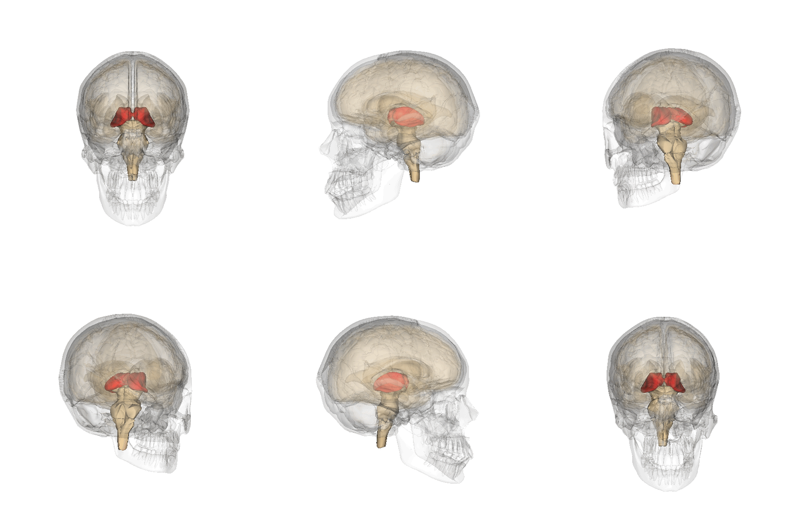
The thalamus is a small, bilateral structure centrally located in the brain. It is highly connected to various brain areas including the hypothalamus, basal ganglia, hippocampus, and more. The thalamus is involved in the regulation of attention, motor coordination, working memory and sleep-wake cycles. In addition, the thalamus regulates sensory information.
The thalamus receives and processes all sensory information except olfactory information. This includes visual, auditory, somatosensory and gustatory information. Essentially, it acts as a gatekeeper, filtering and directing sensory information to the corresponding areas of the cerebral cortex for further interpretation and awareness.
Original by Albert Kok Credits:Wikipedia. CC_BY_3.0
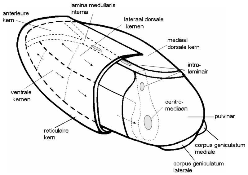
Every nucleus of the thalamus is responsible for the processing of specific information. In humans, the dorsal lateral geniculate nucleus (LGN), medial geniculate nucleus (MGN), ventral posterolateral (VPL) and ventral posteromedial (VPM) are first-order relay nuclei involved in the processing of sensory information.
Original by UC Regents David campus Credits:Wikipedia. CC_BY_3.0
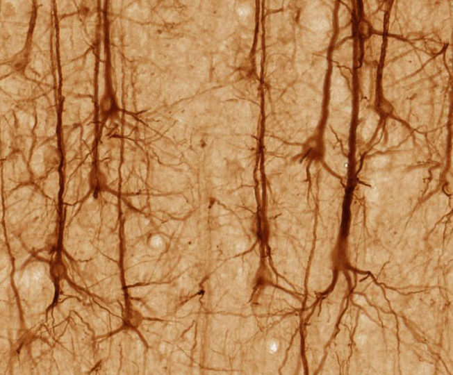
The thalamus consists mostly of excitatory glutamatergic neurons, which relay information to the cortex. Alongside eccitatory neurons, the thalamus also contains inhibitory GABAergic neurons, which control thalamic output by inhibiting excessive activity.
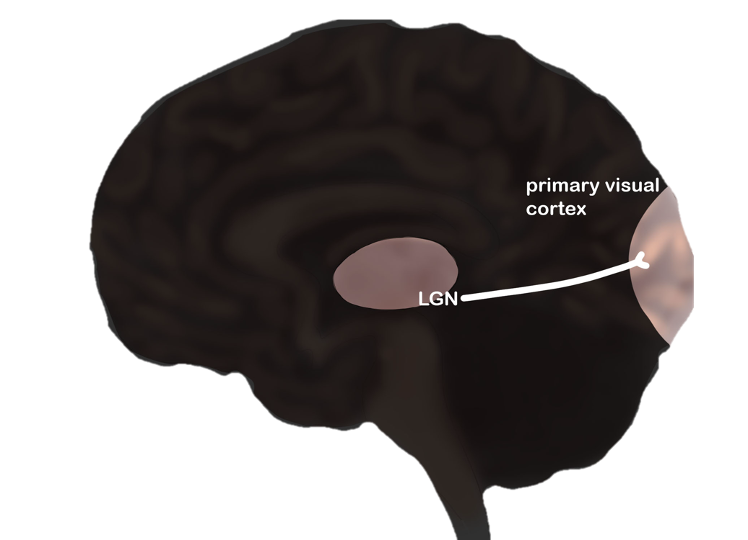
The LGN receives visual input from the retina via retinal ganglion cells and projects this information through relay cells to layer 4 of the visual cortex (Weyand, 2016), located in the occipital lobe. Not only does the visual cortex receive information from the LGN, but it also sends feedback through a descending pathway from layer 6, to modulate the relay cells in the LGN (Weyand, 2016). Notably, ganglion cells in the retina do not exclusively project to the LGN; the proportion of ganglion cells projecting to the LGN varies across species (Weyand, 2016).
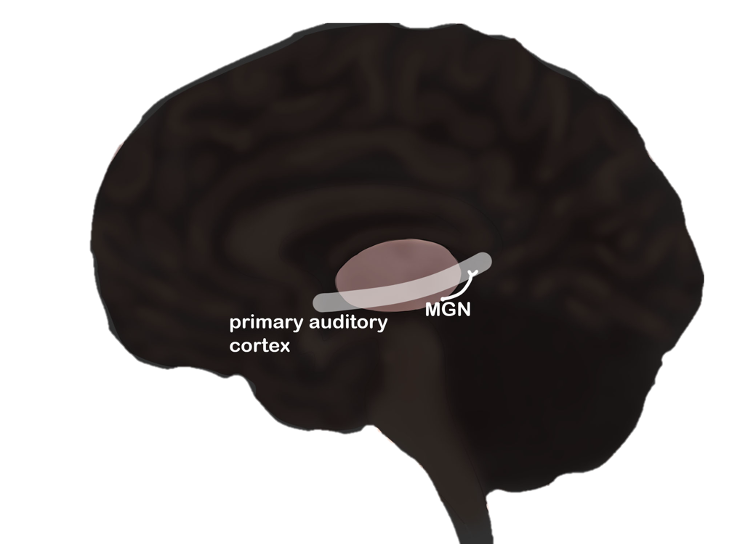
When auditory information reaches the cochlea, it is sent to multiple brainstem centers including the cochlear nuclei (Cant & Benson, 2003 as mentioned in Lee, 2013), present on each side of the brainstem. From this nuclei, auditory information ascends through the inferior colliculus to the MGN (Lee, 2013). Ultimately, the MGN projects this information to the primary auditory cortex (de la Mothe, Blumell, Kajikawa & Hackett, 2006b; Lee & Winter 2008a as mentioned in Lee, 2023) in the temporal lobe.
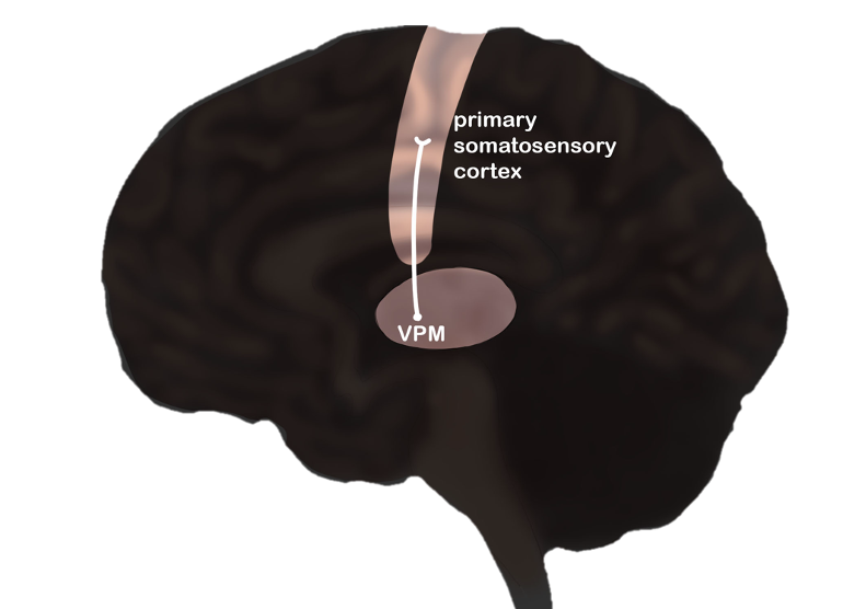
Exteroceptive information received by the face is processed by the VPM (Sheridan & Tadi, 2023). This information is then projected to the primary somatosensory cortex, which is located in the postcentral gyrus of the parietal lobe.
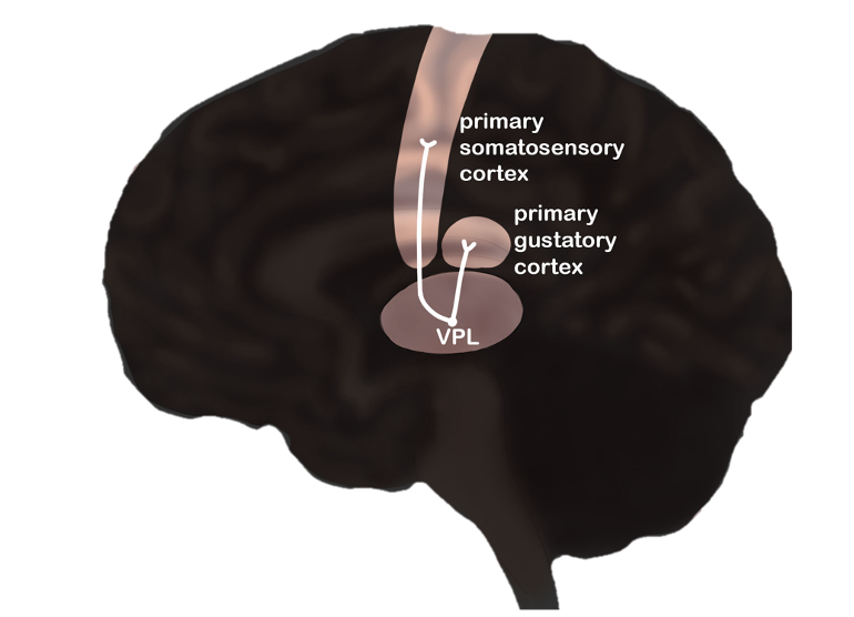
Exteroceptive information received by the body (trunk and limbs) is transmitted by mechanoreceptors, travels through the dorsal column nuclei of the spinal cord and subsequently reaches the VPL (Wang et al., 2002). This information is then projected to the primary somatosensory cortex, which is located in the postcentral gyrus of the parietal lobe. Additionally, the VPL receives gustatory information (Sheridan & Tadi, 2003) via the solitary tract. This information is then relayed to the gustatory cortex, located beneath the frontal and temporal lobes
The thalamic reticular nucleus (TRN)is a thin layer around the thalamus and modulates information flow into and out of the thalamus.
Hinke Boer, Fons Brauers, Andy Louter, Lindsey Pennaertz, Aisha Raja and Tonny Mulder - University of Amsterdam