Brains of this World: Insects
By Tonny Mulder
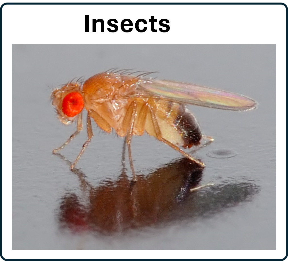
By André Karwath aka Aka; Credits; CC BY-SA 2.5
By Tonny Mulder

By André Karwath aka Aka; Credits; CC BY-SA 2.5
Insects belong to the Phylum Arthropoda, invertebrates with segmented bodies and jointed limbs. Their bodies consist of three segments, the head (A), the thorax (B) and the abdomen (C). The distinctive head and thorax segments seperates them from spiders where the head and thorax are fused into the cephalothorax.
More detailed information can be found here.
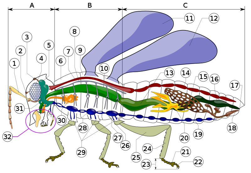
By Piotr Jaworski; Credits; CC BY-SA 3.0
Insect neuroscience is booming with studies on Drosophila being the front runner. Needless to say that databases of Drosophila neuroscience are the first to surface (for an example see below). However, recently the 'Insect Brain Database' was launched and it provides a state of the art database where other insect species are highlighted. Below, several images are presented that were constructed with the 3D brain visualization tool of the database. In these the lateral complex, the central complex and the mushroom bodies in several insect species are visualised. This is just a small selection of insects featured in the insect database, so we strongly encoerage you to visit the insect brain database website. for more indepth and extensive information.
Front page of the insect brain database website.. CC_BY_4.0
The insect brain consists of a number of central brain areas (neuropils), including the circuits of the lateral complex and the central complex that are responsible for navigational control using visual, ideothethic and compass cues and the mushroom bodies that hold long term visual and olfactory memories (Collett and Collett, 2018). This organisation is highly conserved in most insect species, such as the fruit fly (), honey bee (Plath et al., 2017), moths (deVries et al; 2017), locusts (el Jundi et al., 2010) and ants (Habenstein et al., 2020).
Below, the brains of six different insect species are highlighted, featuring the lateral complex (light green), central complex (dark green) and the mushroom bodies (red/orange)
Bogong moth (female)
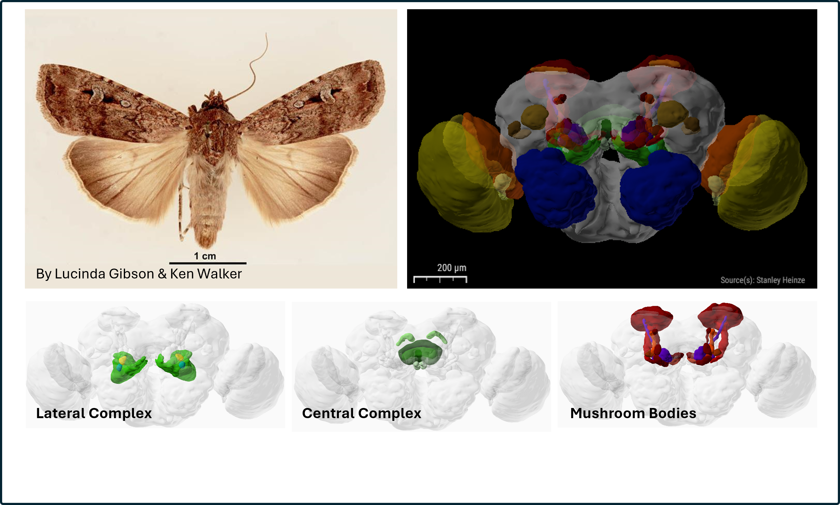
Photo: CC BY 3.0 AU Deed ; insectbraindb (Agrotis infusa): CC_BY_4.0
large earth Bumblebee
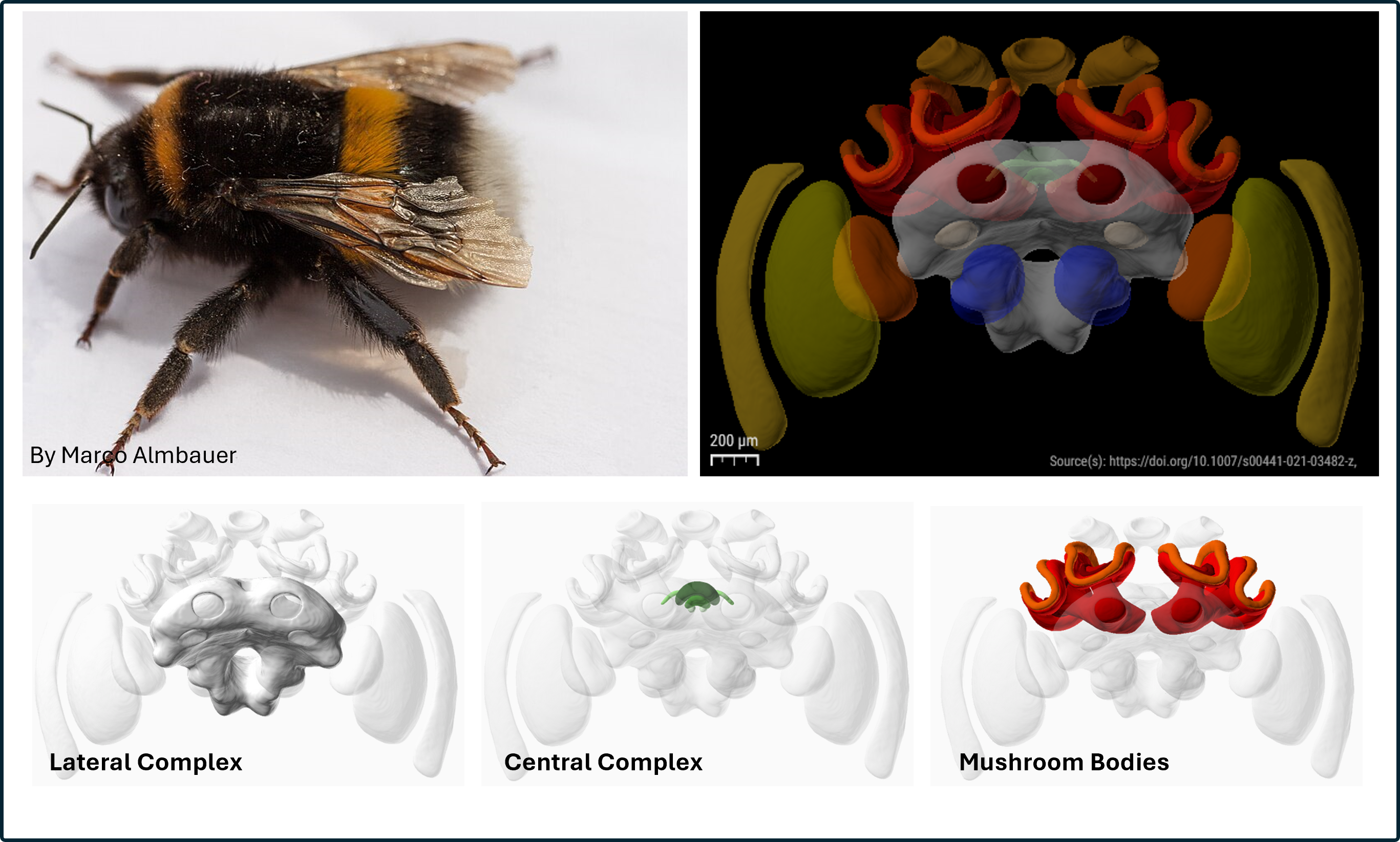
Photo: CC0 1.0 Deed ; insectbraindb (Bombus terestris): CC_BY_4.0
Desert Ant (female)
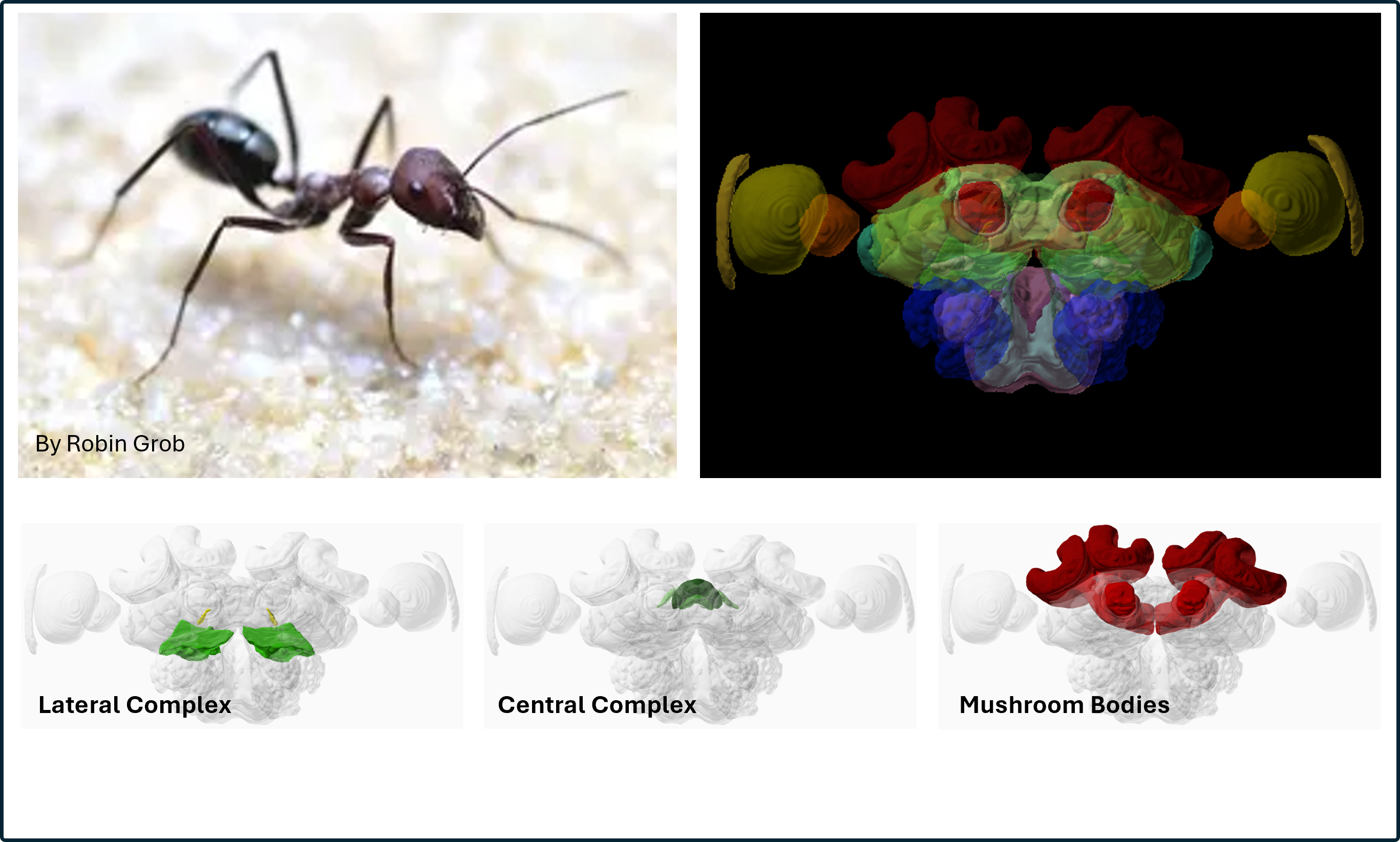
Photo: CC_BY_4.0 ; insectbraindb (Cataglyphis nodus): CC_BY_4.0
Monarch Butterfly (female)
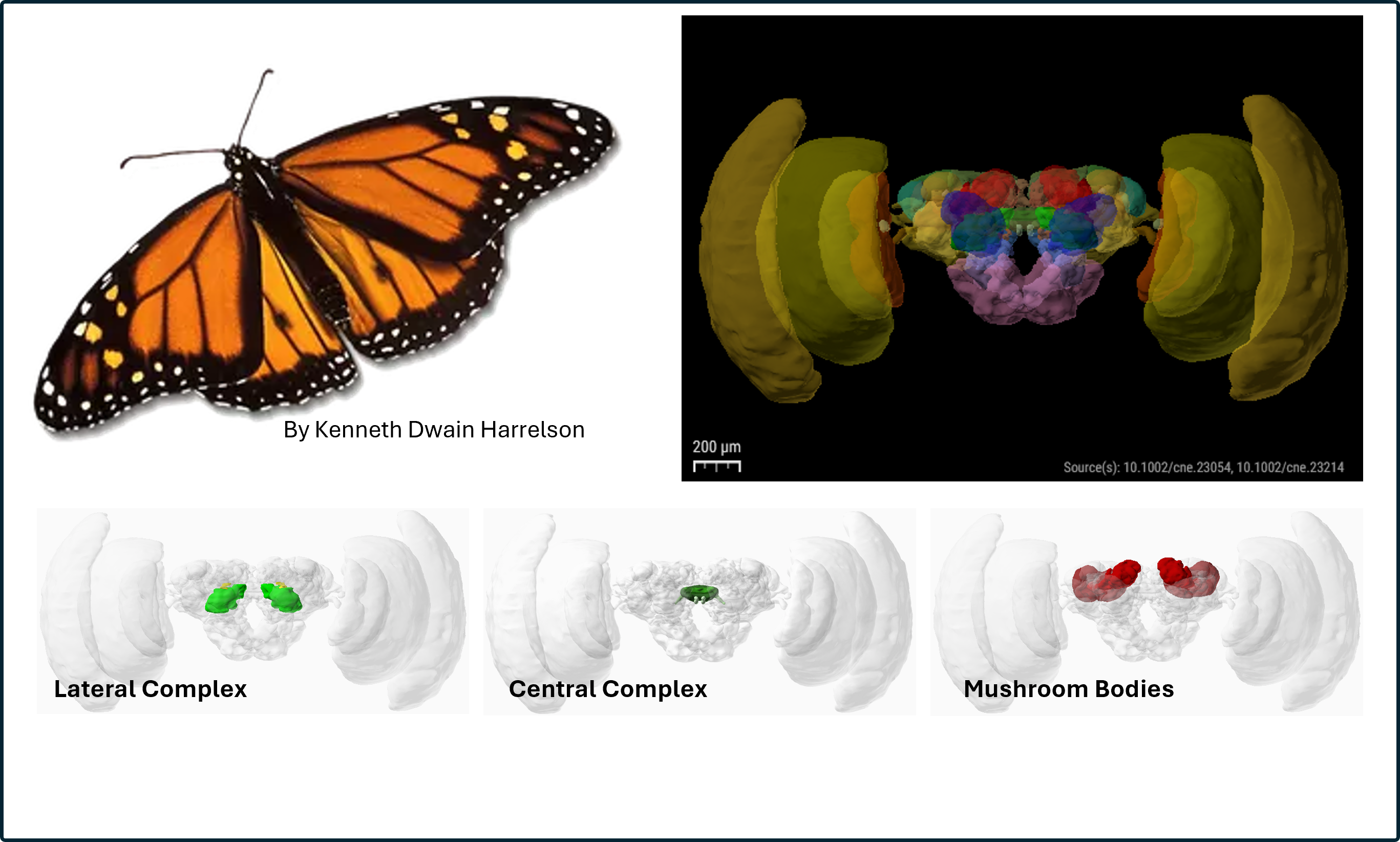
Photo: GFDL ; insectbraindb (Danaus plexippus): CC_BY_4.0
Desert Locust
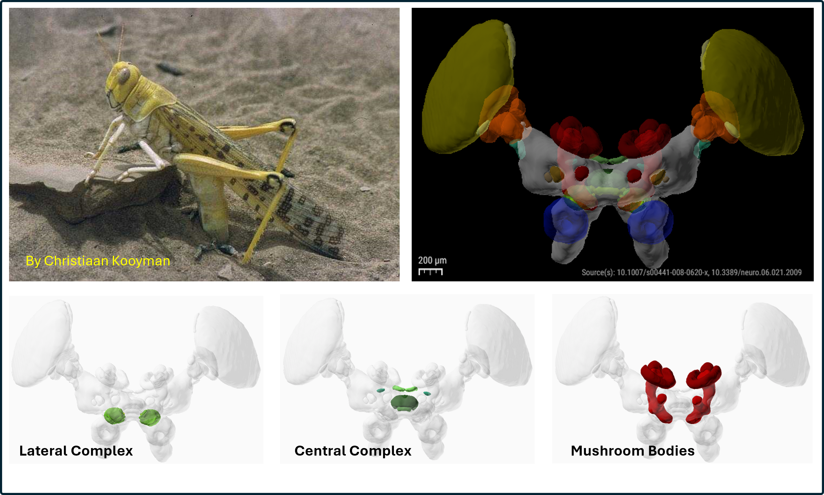
Photo: Public domain ; insectbraindd (Schistocerca gregaria): CC_BY_4.0
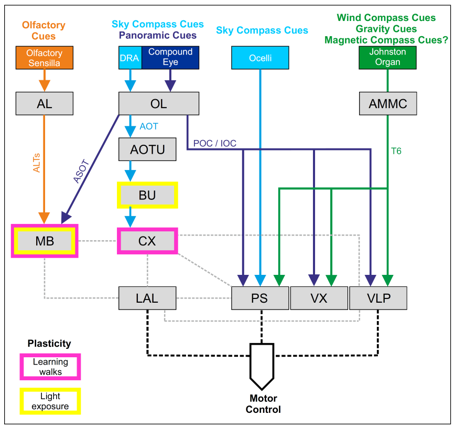
Schematic diagram of brain circuits involved in navigation in the Cataglyphis ants. Rössler et al., 2023.
Legend to picture
lateral Complex
Some More lateral complex

A: Schematic diagram of the brain of an adult Drosophila melanogaster in frontal view. B: The mushroom body in detail. Li et al., 2020. CC_BY_4.0
The Central Complex (CX) consists of a group of unpaired neuropils in the center of the insect brain. The prominent role of the CX is generation of motor outputs according to processed internal and external stimuli (Pfeiffer and Homberg, 2014; Plath and Barron, 2015). As reviewed in Plath et al., 2017, the CX is essential for the initiation and termination of walking, turning and climbing behavior in fruit flies, cockroaches and crickets and is considered as site for action selection and goal-directed behavior. In addition the CX clearly has a role in visual learning and memory involving spatial orientation of the cues in fruit flies and possibly in other insects. Furthermore, the CX might also initiate the appropriate responses to learned stimuli which are processed by the MBs such as color stimuli. Plath et al., 2017.
Central Complex
The brain of Drosophila melanogaster with focus on the central complex

A: Schematic diagram of the brain of an adult Drosophila melanogaster in frontal view, showing the mushroom bodies in color. Li et al., 2020. CC_BY_4.0
B: Subregions of the mushroom body. The alpha, beta and gamma lobes are shown seperately as well as the calex and its subregions. The pedunculus connects the calex complex with the mushroom bodies. Li et al., 2020. CC_BY_4.0
The calex complex comprises of 4 areas:
The brain of Drosophila melanogaster with focus on the sensory inputs to the mushroom bodies
From: Li et al., 2020 CC_BY_4.0
Visual information arrives via the Optic lobe (OL), whereas olfactory and temperature information is acquired via specific sensory receptors in the antennas, processed in the antennal lobe (AL). After being processed in the calex complex, the information reaches the mushroom bodies via the pedunculus. For details visit Li et al., 2020
In early 2023, the 'Virtual Fly Brain' saw daylight (Court et al., 2023). As the authors state in there summary: "The Virtual Fly Brain (virtualflybrain.org) web application and API integrate 3D images of neurons and brain regions, connectomics, transcriptomics and reagent expression data covering the whole CNS in both larva and adult."
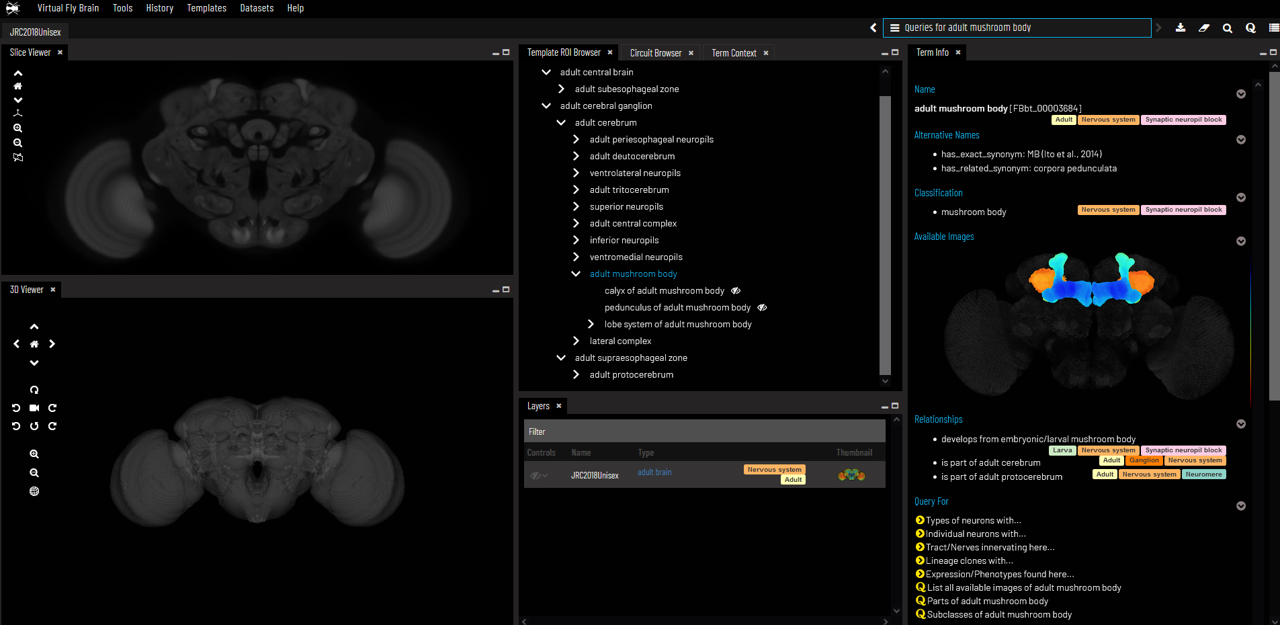
Screen shot of the virtual fly brain website. virtualflybrain.org. CC_BY_4.0
"Users can search for neurons, neuroanatomy and reagents by name, location, or connectivity, via text search, clicking on 3D images, search-by-image, and queries by type (e.g., dopaminergic neuron) or properties (e.g., synaptic input in the antennal lobe). Returned results include cross-registered 3D images that can be explored in linked 2D and 3D browsers or downloaded under open licenses, and extensive descriptions of cell types and regions curated from the literature."
Visual information
Olfactory information
The mushroom body (MB) is a computational center in the drosophila brain. (Lin 2023).
Hinke Boer, Fons Brauers, Andy Louter, Lindsey Pennaertz, Aisha Raja and Tonny Mulder - University of Amsterdam